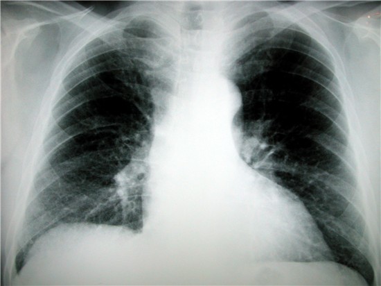
Chest x-ray
A chest X-ray is a picture of your heart, lungs, and chest bones that's made by using a very small amount radiation. Chest X-rays can be used to look for heart and lung abnormalities but cannot alone diagnose angina or coronary heart disease.
This procedure takes about 15 minutes and does not require any preparation1.
Blood tests specifically for cardiac diagnosis are used to measure troponin T (A specific protein released into the blood typically 4-6 hours after heart muscle injury). Cardiac enzymes are also measured in blood samples which show an increase after heart muscle damage. A series of blood tests are taken for both troponin and cardiac enzymes which indicate the extent of heart muscle damage.
It is important to note that troponin levels may remain high for 1 to 2 weeks after a heart attack.
Increased troponin levels may also be due to:
- Abnormally fast heart beat.
- High blood pressure in lung arteries (pulmonary hypertension).
- Blockage of a lung artery by a blood clot, fat, or tumor cells (pulmonary embolus).
- Congestive heart failure.
- Coronary artery spasm.
- Inflammation of the heart muscle usually due to a virus (myocarditis).
- Strenuous exercise (for example, due to marathons or triathlons).
- Trauma that injures the heart such as a car accident.
- Weakening of the heart muscle (cardiomyopathy).
Increased troponin levels may also result from certain medical procedures such as:
-
Cardiac angioplasty/stenting.
-
Heart defibrillation or electrical cardioversion.
-
Open heart surgery.
-
Radiofrequency ablation of the heart2.
Electrocardiogram (ECG)
An ECG is a non-invasive test which takes a reading of the electrical activity of your heart and records the strength and timing of electrical signals as they pass through each region of the heart. An ECG can show evidence of heart damage due to coronary heart disease, signs of a previous or current heart attack, abnormal rhythms, unstable angina, congenital heart abnormalities, abnormal electrolytes and inflammation of the heart muscle. 10 electrode stickers and leads are placed on the skin around the left side of the chest wall, arms and legs. You will be asked to lie still while a reading is taken which takes approximately 10 minutes.
Exercise Stress Test
An exercise stress test involves an ECG being performed during exercise, commonly on a treadmill, to test how your heart tolerates exercise.
An Exercise Stress Test measures and records your heart's electrical activity whilst under the strain of exercise; walking on a treadmill (If you are unable to walk on a treadmill the Doctors may choose to increase your heart rate with medication and the procedure is performed lying down). The treadmill will increase in speed and slope at specific intervals and your heart rate, blood pressure and electrocardiogram (ECG) will be continually monitored. A cardiologist and a cardiac technician will be present at all times. This test takes approximately 30 minutes. Comfortable footwear and clothing is the only preparation required3.
24 hour Blood Pressure Monitoring
A blood pressure monitor records your blood pressure at intervals throughout the day and night.
The monitor itself is a small box approximately 10x7x3cm in dimension. The monitor is worn on a belt around your waist and there will be a tube connecting the box to a cuff which is worn on your least dominant arm. The monitor automatically inflates every 20 minutes until 10pm, then every hour until 6am the following day and then again every 20 minutes until the monitor is removed. When the cuff begins to inflate you are required to keep your arm relaxed and still down by your side until the cuff deflates. The cuff will feel very tight as the pressure rises. During the recording you will need to list the time that the cuff inflates and your activity at that time. On completion the device is removed by a technician, the data is downloaded and then reported on by a cardiologist4.
Holter Monitor
A holter monitor records your heart rhythm continuously throughout the day and night.
The monitor can be worn anywhere from 1-7 days which is determined by the cardiologist. It is the size of a mobile phone and consists of 5 or 7 leads which are attached to your chest by adhesive electrodes with tape placed over the top to ensure they remain in place. The holter will either be hung loosely around your neck by a strap or clipped onto your waistband. During the recording you will need to list your symptoms, the time they occur and what you were doing at the time on the diary sheet provided. Normal daily activities can be carried out BUT the monitor must remain dry i.e. no swimming or showering, nor use an electric blanket or magnetic underlay.5
Echocardiogram
An Echocardiogram (or “Echo”) is an ultrasound of your heart. It provides essential information about the structure and function of your heart chambers, valves and related vessels. This test takes approximately 40 minutes. A small probe will be placed over different areas of your chest in order to assess the heart from multiple views. A water soluble gel is used as a contact medium between the probe and your chest wall to improve image quality.
You will be able to see images of your heart on the screen and hear noises of blood flow. You will feel pressure on your chest where the sonographer is imaging. A series of standard measurements will be taken throughout the test. All images are stored digitally and reported by a cardiologist6.
Coronary Angiogram
Coronary angiography is a procedure in which a special x-ray is taken of your heart’s arteries.
This investigation examines your coronary arteries to see if they are narrowed or blocked. It is performed when your doctor suspects or knows that you have coronary heart disease.
A local anaesthetic is administered into the groin, wrist or elbow then a catheter (a long thin tube) is inserted into the artery. The catheter is moved up the inside of your artery until it reaches your heart. A special dye is injected into your coronary arteries and x-rays are taken. The x-ray gives detailed information about the state of your heart and coronary arteries assisting doctors in determining what treatment will be best7.
The catheter is removed and the radiologist will apply pressure on the artery for at least ten minutes. You must then lie flat for about three hours to let the artery seal up firmly. You will then be allowed to sit up and should avoid strenuous activities for the next 12–24 hours. This investigation will require Hospital admission8.
Computer Imaging
Computer imaging (tomography) refers to diagnostic imaging tests that use computer aided techniques to gather images of the heart. They include CT, PET and MRI scans. This imaging can evaluate aortic disease, cardiac masses and pericardial disease.
CT (Computerised Tomography)
CT is a non-invasive test that uses x-rays to take 3D images of the heart. Images can be taken of the beating heart, and show calcium and blockages in the heart arteries. Pictures can be created of the healthy and diseased parts of your heart. This investigation can assist in finding problems in your heart’s structure and in how your heart pumps blood, blockages, scarring from heart attack, fluid in the pericardial sac (the lining over the heart surface), plaque build-up and abnormalities in the large vessels leaving your heart.
Contrast dye is sometimes used for this test. It is important for your Doctor to know if you are allergic to dyes, iodine or shellfish. Check with your Doctor what period of fasting is required.
During the test you lie down on a table with ECG electrodes attached to your chest to monitor your ECG. The table slowly moves inside the machine. If a contrast dye is used, it is injected through an intravenous line (IV) placed in an arm vein.
You can talk to the technician via a two-way intercom and you will be asked to hold your breath for short periods. The process takes 5-10 minutes10.
PET scan (Positron Emission Tomography)
A PET scan of the heart is a non-invasive nuclear imaging test using radioactive tracers to produce pictures of your heart. Radioactive tracers mix with the blood and are taken up by the heart muscle and a special “gamma” detector that circles the chest picks up signals from the tracer. A computer converts the signals into pictures of your heart at work. Doctors use cardiac PET scans to diagnose coronary artery disease (CAD) and heart damage. These scans can show healthy and damaged heart muscle and can help find out if you will benefit from a percutaneous coronary intervention (PCI) such as angioplasty and stenting, coronary artery bypass surgery (CABG) or another procedure.
This investigation is performed at a Hospital. You will need to fast prior to this procedure, your medications may need to be withheld and your caffeine intake altered.
ECG electrodes will be placed on your chest and the tracer will be injected through an IV line into your arm. You will lie on a flat table that’s connected to the PET scanner and a computer. The table will slide into the scanner, which is shaped like a giant doughnut.
Within the PET scanner, detectors record the radioactive patterns of the tracer in your heart. The information is transformed into images on a computer screen.
Several scans are done over time to provide pictures of thin slices of your entire heart from all angles. It’s very important to hold completely still with your arms above your head while each scan is being done10.
MUGA (multiple-gated acquisition)
A MUGA scan is a type of nuclear imaging test which shows how well your heart is pumping.
A radioactive tracer (called a radionuclide) and a special camera is used to take pictures of your heart as it pumps blood. The test measures how well your heart pumps with every heartbeat and may be done while you stay still, exercise or both. It measures your ejection fraction, which is the amount of blood pumped out of the heart during each heartbeat (contraction) and is expressed as a percentage. For example, an ejection fraction of 60 percent means that 60 percent of the total amount of blood in the left ventricle when it is full is pumped out with each heartbeat. A normal ejection fraction is between 50 and 75%.
During this scan, a small amount of a radioactive substance or tracer (called a radionuclide) is injected into your blood and excreted through the kidneys within 24 hours. The tracer attaches to your red blood cells and a gamma camera takes pictures of your heart. The pictures show if areas of your heart muscle aren’t contracting normally and show how well your heart pumps blood. You may be asked to fast or withhold caffeine, with hold some medication and wear comfortable, clothing and comfortable shoes prior to this test.
This scan is performed in a Hospital. ECG Electrodes are placed on the chest, radionuclide is injected intravenously and a camera will take many pictures of your heart while you’re resting or exercising which takes 1-2 hours11,12.
Magnetic Resonance Imaging (MRI)
Magnetic Resonance Imaging (MRI) is a method of imaging which utilises a strong magnetic field to generate images of any region of the human body. A cardiac MRI is used by doctors to assist in the diagnosis and treatment of heart disease. A signal is detected by an antenna or small coil which is placed around the body part being scanned. This signal is encoded by magnetic field gradients which are responsible for the loud noise associated with MRI. The signal is mathematically processed by powerful computers to form an image. MRI scanners are generally about 1.8m in length, shaped like a long tube and open at both ends. Some MRI examinations require an injection of a contrast medium into an arm vein to allow imaging of blood vessels/vascular tissues.
MRI is not suitable for patients with pacemakers and various other medically implanted devices due to the strong magnetic fields13.
TOE (Transoesophageal Echocardiogram)
A trans-oesophageal echocardiogram (T.O.E), is similar to an echocardiogram, but is performed via an ultrasound transducer, or probe, sitting in the oesophagus or food pipe. This test takes approximately 40 minutes. The T.O.E. probe will lie much closer to the heart than a traditional probe on the chest wall, providing a clearer and more detailed view of your heart’s structures and can check for clots (thrombus) in the atria.
This test requires you to fast for 6 hours, a small dose of relaxant is given intravenously, a probe will be passed over the tongue into the back of the throat and at this point you will be asked to swallow. An effective swallow will have the probe in the correct position so that you can relax while the images are being acquired you will be observed for a short time after the test and then allowed to go home accompanied by another adult14.